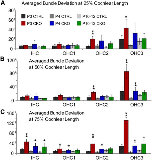
Figure 6.
The extent of planar polarity refinement is graded along the length of the cochlea. A–C, Averaged stereociliary bundle deviations from the mediolateral axis for hair cells located at the 25% (A), 50% (B), and 75% (C) analysis fields from tissues collected at three stages of postnatal development. Planar polarity refinement actively reorients stereociliary bundles in the Vangl2 CKO between P0 and P4, whereas bundle polarity does not change significantly between tissues collected at P4 and P10–P12. At all ages, the differences in averaged stereociliary bundle deviation from the mediolateral axis are greatest in the most apically positioned 75% analysis field (C). Despite planar polarity refinement, IHCs and hair cells located in OHC3 remain modestly misoriented and are never as organized as the corresponding hair cells in littermate controls. The number of mice assayed at P0 is n = 4 (25%), 4 (50%), 4 (75%) for WT and n = 6, 6, 6 for CKO; at P4, n = 3, 3, 3 for WT and n = 5, 6, 6 for CKO; and at P10–P12, n = 2, 4, 6 for WT and n = 4, 4, 6 for CKO. Error bars show SD, and asterisks indicate statistical significance for differences between Vangl2 CKOs and littermate controls of the same hair cell type and cochlear position calculated using two-tailed Student's t test with unequal variance (*p < 0.05, **p < 0.005).

