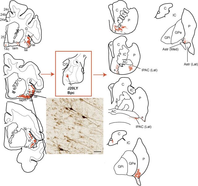
Figure 5.
Distribution of retrogradely labeled cells in the insula cortex (left), and anterogradely labeled fibers (right) in the striatum resulting from an injection in the rostromedial Bpc (J20LY). This region of the amygdala received input from all subdivisions of the insula (rostral to caudal is displayed top to bottom). Except for a few retrogradely labeled cells in area 14c, no retrogradely labeled cells were found in any other PFC region. Insert photo shows cells labeled with LY tracer in Ial <20×, bright-field microscopy. Scale bar, 50 μm. Striatal output targets involved the rostromedial shell (s) and core, lateral IPAC, rostroventral caudate body, and caudoventral putamen. AC, anterior commissure; C, caudate nucleus, G, gustatory cortex; GPe, globus pallidus, external segment; GPi, globus pallidus, internal segment; IC, internal capsule; OT, optic tract; P, putamen; V, ventricle.

