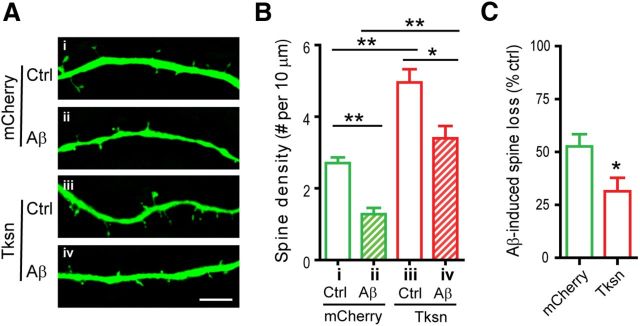
Figure 2.
Forced expression of α1-takusan alleviates Aβ-induced loss of dendritic spines in cultured neurons. A, Cultured hippocampal neurons were infected with SFV encoding mCherry (i, ii) or mCherry-α1-Tksn (iii, iv) and then exposed to Ctrl (i, iii) or Aβ CM (ii, iv). Dendritic spines were visualized by EGFP expressed from a lentiviral vector. Scale bar, 5 μm. B, The density of dendritic spines was significantly decreased by Aβ-application in both mCherry-expressing cells (ii vs i) and Tksn-expressing cells (iv vs iii), and was increased by Tksn overexpression (iii vs i, and iv vs ii). Values (clusters per 10 μm) for the four conditions are as follows; i, 2.70 ± 0.16; ii, 1.28 ± 0.17; iii, 4.96 ± 0.37; iv, 3.40 ± 0.35. C, Aβ-induced synaptic loss in the density of dendritic spines (percentage control) is significantly less in the cells overexpressing mCherry-α1-takusan than in the cells overexpressing mCherry (31 vs 52%, p < 0.05). Values are mean ± SEM. n = 10. *p < 0.05, **p < 0.01, one-way ANOVA or Student's t test.

