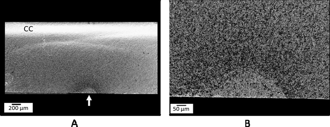
Figure 1.
Fracture surface of a specimen from Group 3 (mechanical aging) evidencing the presence of a slow crack growth halo. (A) General view - the white arrow point to the critical flaw that is surrounded by a darker halo (SCG halo), and it is also possible to observe a compression curl in the opposite side (CC). (B) Close view of the critical flaw and SCG halo.

