Figure 2.
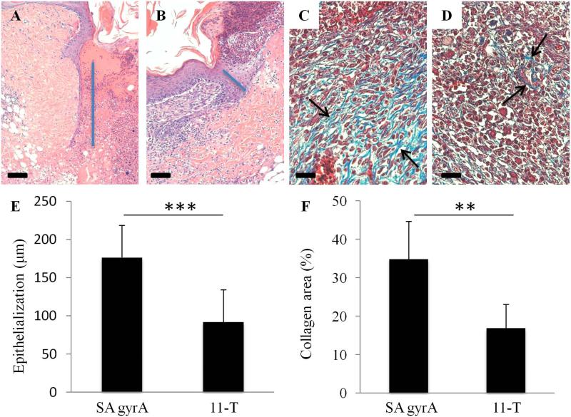
Conjugate drug treated wounds show improved epithelialization and increased mature collagen. The increased epithelial tongue (blue bar) length at 10 days shows improved migration and wound closure in treated (A) wounds when compared to control (B) wounds. There is a statistically significant increase in epithelial tongue length in wounds that received the drug (E, p < 0.001, average + SD). Trichrome stain showed a significantly increased amount of mature collagen (highlighted by arrows), as indicated by a light blue stain, in conjugate treated tissue (C). S. aureus infected wounds that received inactive conjugate (D) showed minimal mature collagen at day 10. Quantification of collagen positive area per high power field (F) confirmed the increase in collagen (p < 0.01). Means compared using Student's t-test. Scale bar is 100 μm in A and B and 50 μm in C and D.

