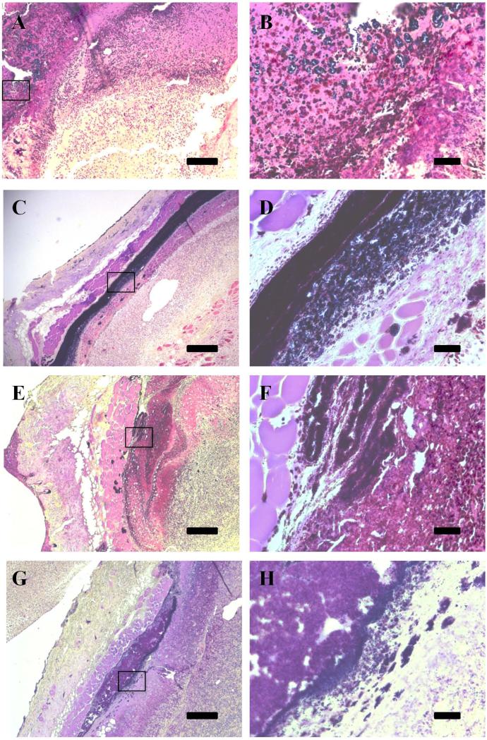
Figure 3.
Gram stain shows reduced bacterial load in wounds receiving active conjugate. Treated wounds at 7 (A,B) and 10 days (E, F) show decreased gram positive staining when compared to control wounds at 7 (C, D) and 10 (G, H) days. Inset images (B, D, F, and H) highlight areas in the tissue where staphylococcal colonies are visible in the tissue. Scale is 200 μm for A, C, E, and G, and is 25 μm for B, D, F, and H.

