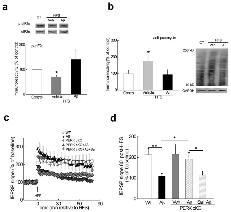
Figure 2. Aβ-induced impairment in LTP is alleviated by deleting the eIF2α kinase PERK.
(a) LTP-inducing stimulation decreased the phosphorylation of eIF2α (middle lane), which was reversed by Aβ(1–42) (right lane). Slices were harvested 30 minutes post-HFS and area CA1 was microdissected for Western blot analysis. n=8. *p<0.05. (b) De novo protein synthesis (assayed by SUnSET). LTP-inducing stimulation increased de novo protein synthesis, which was blunted by Aβ(1–42). Slices were harvested 30 minutes post-HFS and area CA1 was microdissected. n=4. *p<0.05. (c) Treatment of hippocampal slices from WT mice with 500 nM Aβ(1–42) resulted in impaired LTP (grey triangles, n=8) compared with LTP in vehicle-treated WT slices (open squares, n=7). In contrast, LTP was induced in slices from PERK cKO mice in the presence of Aβ(1–42) (half-filled triangles, n=6), which was blunted by application of 10 μM Sal003 (Sal, grey diamonds, n=7). In addition, HFS induced LTP in PERK cKO mice that was comparable to that in WT littermates (grey circles, n=7). (d) Cumulative data showing the mean fEPSP slope 80 min post-HFS from the LTP experiments in panel c. *p<0.05; **p<0.01. Full-length blots/gels are presented in Supplementary Figure 7.

