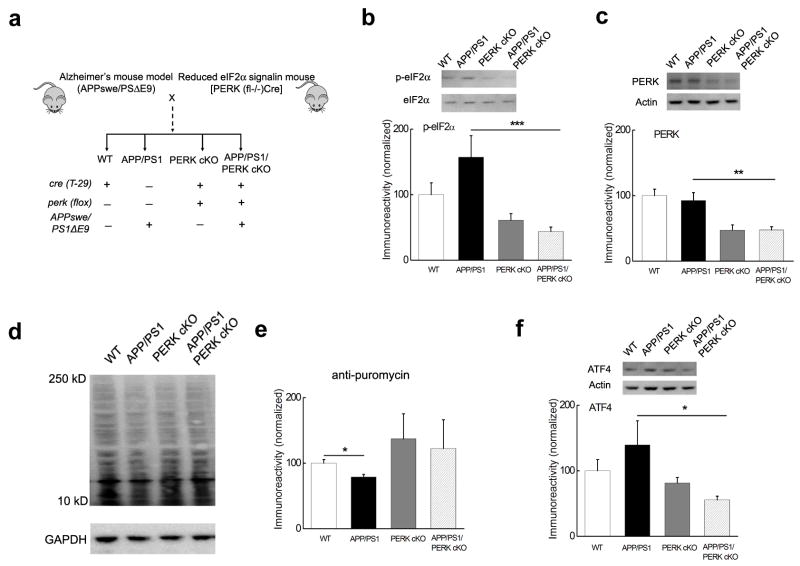
Figure 3. Generation of AD model mice with reduced PERK-eIF2α signaling.
(a) Diagram depicting the creation of mice with AD-associated transgenes and reduced PERK/eIF2α signaling. (b) eIF2α phosphorylation was reduced in hippocampal area CA1 of APP/PS1/PERK cKO mice compared to the increased levels of eIF2α phosphosphorylation in APP/PS1 mice, which was correlated with the expression of PERK (c). n=10 for APP/PS1/PERK cKO, n=6 the other three groups. (d) Representative Western blot showing that de novo protein synthesis (assayed by SUnSET) was reduced in APP/PS1 mice compared to WT littermates. In addition, de novo protein synthesis in PERK cKO and APP/PERK cKO mice was not different from WT mice. (e) Cumulative data showing densitometric analysis of experiments in panel d. n=4. *p<0.05. (f) Elevated levels of ATF4 in APP/PS1 mice were reduced to WT levels in APP/PS1/PERK cKO mice. Western blots were performed on tissue from area CA1 of the hippocampus. n=10. All data for the densitometric analysis of the Western blots were presented as mean ± SEM. *p< 0.05, **p< 0.01, ***p<0.001. Full-length blots/gels are presented in Supplementary Figure 7.

