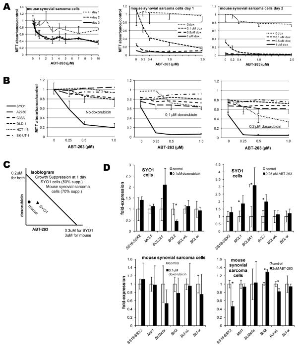
Figure 3. ABT-263 inhibits synovial sarcoma cell lines in vitro.
(A) Increasing concentrations of ABT-263 as a single agent modestly decrease proliferation/survival of mouse synovial sarcoma cells in vitro as determined by a proliferation assay, but only meaningfully by 2 days following application. Even nanomolar concentrations of ABT-263 enhanced the cytotoxicity of doxorubicin in these mouse synovial sarcoma cells. (B) The SYO1 human synovial sarcoma cell line displayed marked sensitivity to nanomolar concentrations of ABT-263 as single agent or in combination with doxorubicin in proliferations assays performed 1 day after drug application. Side-by-side comparison to 5 other human cancer cell lines demonstrated their poor sensitivity to ABT-263 as a single agent or in combination with doxorubicin, except for HCT116, a colon cancer cell line. (C) Isobologram for 50% suppression of growth and proliferation of SYO1 cells after one day of ABT-263 and/or doxorubicin and 70% suppression of mouse synovial sarcoma cells demonstrate synergism between these drugs for both cell lines. (D) Most changes in gene expression following application of doxorubicin or ABT-263 to SYO1 cells or mouse synovial sarcoma cells are not significant. †While the expression of BCL2A1 increases significantly following application of ABT-263 to SYO1 cells, it remains at a barely detectable level. Each proliferation assay had a sample size of 8 in each drug concentration at each time point following application, expression following drug applications had 6 replicates. All results are presented as means with standard deviation error bars. Asterisks denote p-values < 0.05 by Student’s t-test.

