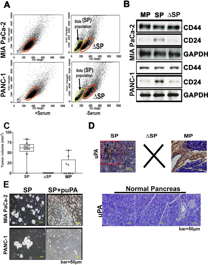
FIGURE 1:
Stem cell–like properties of the SP cells derived from pancreatic cancer cells. (A) Mixed populations of MIA PaCa-2 and PANC-1 cells (2 × 10⁶) were sorted by density-based flow cytometry (10,000 cells sorted per treatment condition, with three replications) to separate SP and ΔSP cells. Acquisition was performed on a FACSCalibur flow cytometer, and viable cells were analyzed with CellQuest software. (B) Cell lysates prepare from the sorted SP and ΔSP cells were immunoblotted for CD24 and CD44 to elucidate expression of cancer stem cell markers. (C) SP, ΔSP, and MP cells were implanted subcutaneously in nude mice (10,000 cells/mouse), and the tumor volumes in treated groups were quantified and represented graphically (mean ± SD; n = 5 and p < 0.001). (D) Subcutaneous tumors grown as in C were implanted orthotopically in the pancreas of nude mice as described in Materials and Methods and allowed to grow for 40 d. At the end of this period, pancreatic tissues were harvested and processed for paraffin sectioning. Expression levels of uPA were determined by immunohistochemistry using anti-uPA and control immunoglobulin G. Brown color denotes uPA-antibody–positive reaction. Normal pancreatic tissue was also sectioned and immunoprobed for uPA. (E) Proliferation and formation of the neurospheres by untreated SP cells derived from MIA-PA Ca-2 and PANC-1 cells (left). Right, disintegration of the neurospheres after exposure to shRNA specific for uPA (puPA).

