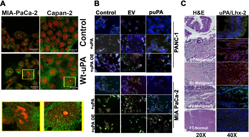
FIGURE 3:
Nuclear uPA regulates expression of Lhx2 in pancreatic cancer cells. (A) MIA-PaCa-2 and Capan-2 cells were left untreated or incubated with 20 nM of recombinant WT-uPA for 1 h, fixed in MeOH, and stained with anti-uPA rabbit polyclonal Abs and Alexa 488–conjugated anti-rabbit secondary Abs. Nuclei were counterstained with propidium iodide (red). Green staining denotes cytoplasmic and nuclear localization of uPA. (B) MIA PaCa-2 and PANC-1 cells plated and grown on chamber slides were transfected with pSV (scrambled vector) or puPA to lower uPA or uPA-encoding plasmid for uPA overexpression (pUPAOE). Nontransfected cells were also incubated with exogenously added WT-uPA protein. Cells were immunoprobed for uPA (green) and Lhx2 (red) and mounted with DAPI-containing mounting medium, and fluorescent photomicrographs were obtained as described (Stepanova et al., 2008). (C) Human pancreatic cancer tissue array (± cancer) was stained with H&E or immunoprobed for uPA or Lhx2 (A1, C1, E1, and F2 are malignant pancreatic adenocarcinoma tissues, and F7 is normal pancreatic tissue).

