
FIGURE 5:
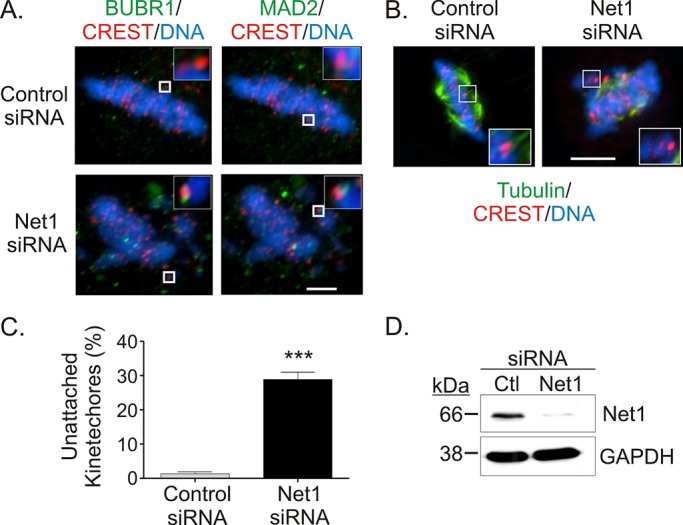
Spindle assembly checkpoint activation and reduced spindle stability in Net1-depleted cells. (A) HeLa cells were transfected with control or Net1-specific siRNAs. Two days later the cells were fixed and stained for the spindle assembly checkpoint proteins BubRI (left, green) or Mad2 (right, green), CREST (red), and DNA (blue). Spindle assembly checkpoint activation was determined by adjacent localization of BubRI or Mad2 with CREST in metaphase cells (insets). Representative micrographs. Bar, 2 μm. (B) Control or Net1 siRNA–transfected cells were incubated at 4°C for 10 min before fixation. The cells were then stained for α-tubulin (green), CREST (red), and DNA (blue). Representative micrographs. Bar, 10 μm. (C) Quantification of unattached kinetochores in control or Net1 siRNA–transfected cells after cold treatment. Average of 25–30 kinetochores counted/cell from three independent experiments. Errors are SEM. Statistical significance was determined by Student's t test; **p < 0.001. (D) Representative Western blot of siRNA-transfected cells.