
FIGURE 3:
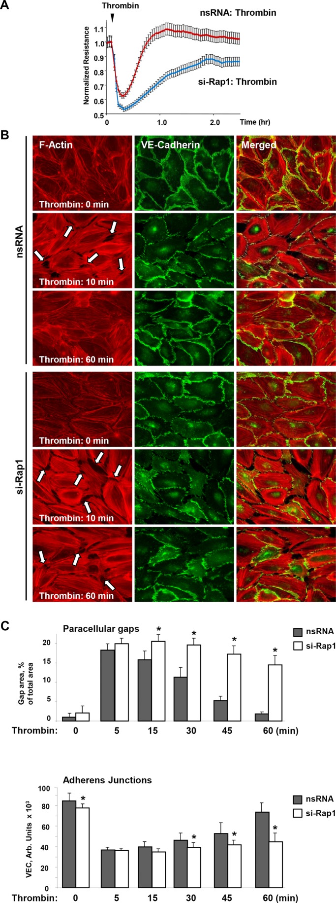
Effects of Rap1 knockdown on functional and structural barrier restoration of pulmonary EC monolayer after thrombin. (A) ECs plated on microelectrodes were transfected with Rap1-specific siRNA or nonspecific RNA 48 h prior to TER measurements. Control and Rap1-specific siRNA-treated ECs were stimulated with thrombin at the time indicated by the arrow, and TER changes were monitored over time. (B) ECs plated on glass coverslips were transfected with Rap1-specific siRNA or nonspecific RNA prior to stimulation with thrombin. Immunofluorescence staining of F-actin (left) and VE-cadherin (middle) was performed using Texas Red phalloidin and VE-cadherin specific antibody, respectively. Right, merged images of F-actin and VE-cadherin staining. Arrows indicate areas of thrombin-induced intercellular gap formation. (C) Quantitative analysis of gap formation and cell junction VE-cadherin localization in control and Rap1-depleted ECs at different times after thrombin treatment. Data are expressed as mean ± SD; *, p < 0.05.