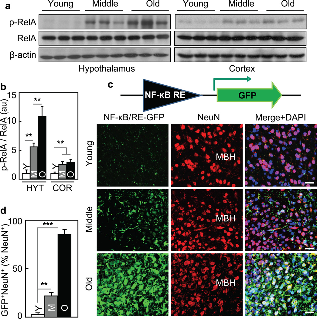
Figure 1. Aging-dependent hypothalamic NF-κB activation.
57BL/6 mice (chow-fed males) were analyzed at young (3–4 months) age (Y), middle-old (11–13 months) age (M), and old (22–24 months) age (O). a&b. Hypothalami were analyzed via Western blots. b: Intensity of p-RelA normalized by RelA (au: arbitrary unit). c&d. Mice received MBH injections of lentiviral GFP controlled by NF-κB response element (NF-κB/RE), and following ~3-week recovery, brain sections were made to reveal GFP and NeuN stainining. DAPI staining shows entire cell populations. Bar = 25 µm. d: Percentages of cells co-expressing GFP and NeuN (GFP+NeuN+) among NeuN-expressing cells (NeuN+) in the MBH. **P < 0.01, ***P < 0.001; n = 6 (b) and 3 (d) per group. Error bars reflect mean ± SEM.

