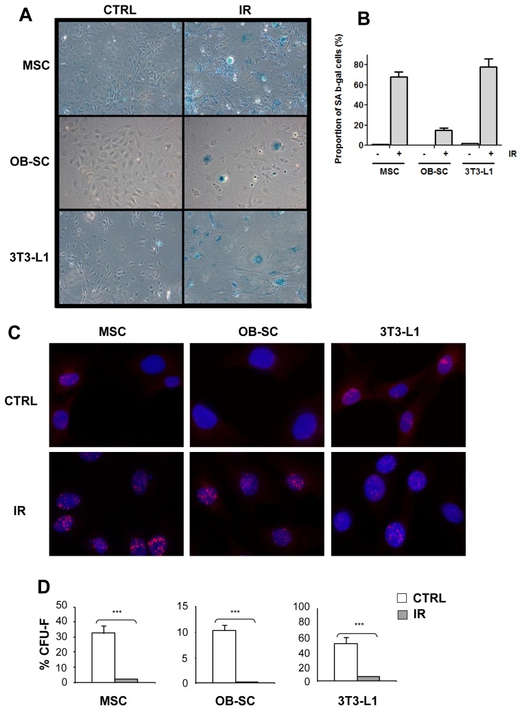
Figure 1. Senescence of multipotent and committed stromal lineages following exposure to IR.
(A) Murine bone marrow-derived multipotent stromal cells (MSC), osteoblasts (OB–SC) and pre-adiopocytes (3T3-L1) were exposed (IR) or not (CTRL) to 10 Gy IR and 7 days later stained for the expression of the senescence-associated β-galactosidase (SAβ-gal). (B) Quantification of the proportion of SAβ-gal positive cells in each population. (C) Sustained activation of the DNA damage response in stromal populations was measured by staining for the presence of 53BP1 DNA damage foci (in red) one week post exposure to IR. Nuclei were counterstained with DAPI. (D) The proliferation capacity of MSC, OB–SC and 3T3-L1 cell population was determined using a CFU assay one week post-exposure or not to IR. Mean ± standard error of at least three individual experiments is shown. p values were obtained by performing a Student’s t-test.

