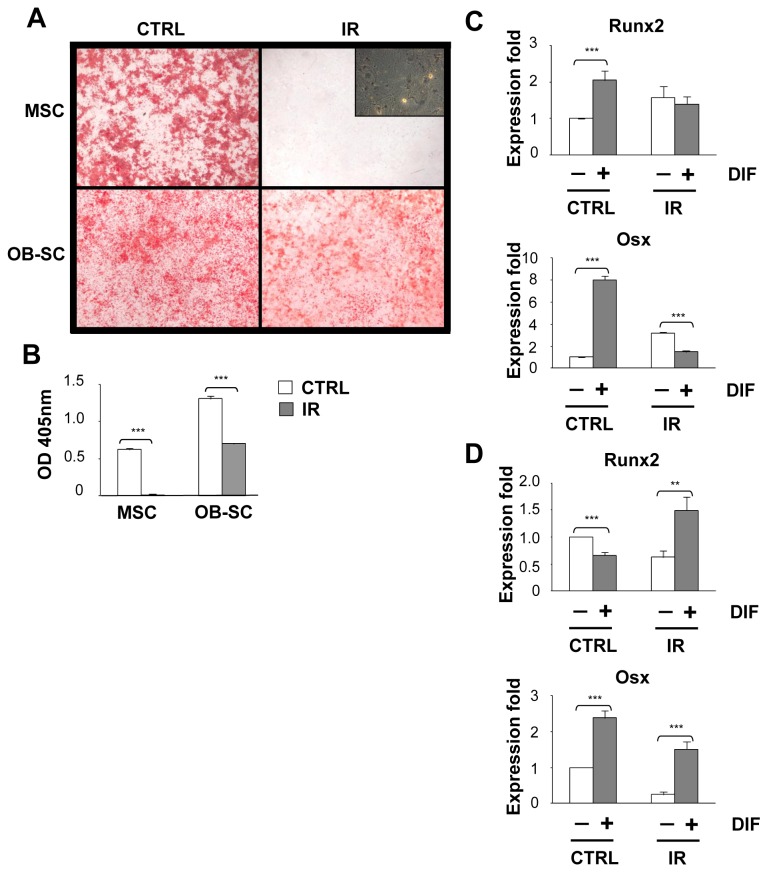
Figure 3. Abrogation of osteogenic differentiation potential following irradiation is limited to stromal progenitor cells.
(A) MSC and osteoblasts (OB–SC) were exposed (IR) or not (CTRL) to 10 Gy IR and one week later placed in osteogenic differentiation media for 14 to 21 days. Representative photographs showing mineralization nodules accumulation stained with Alizarin Red S is shown for each population. Scale bar: 2mm. Phase contrast photograph showing the presence of senescent MSC in absence of mineralization is also shown. (B) Quantification of mineralization was determined by the extraction of Alizarin Red S and detection by spectrophotometry. (C and D) Expression of Runx2 and Osx was determined by quantitative real-time PCR using RNA extracted from control and IR-induced senescent MSC and OB–SC populations cultured or not in osteogenic differentiation media. Mean ± standard error; *: p value < 0.05.

