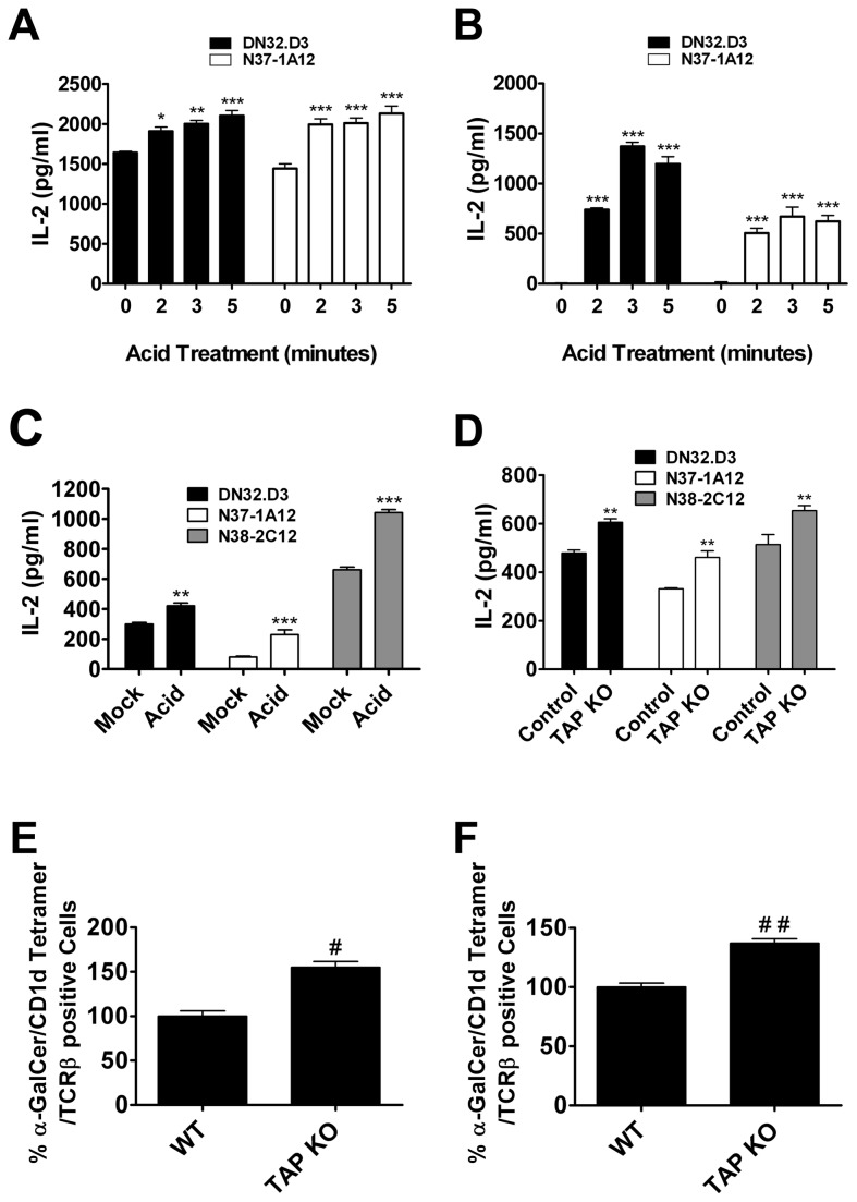
Figure 1. Disruption of cell surface MHC I molecules enhances CD1d-mediated Ag presentation.
(A–C) Antigen presenting cells (A) LMTK-CD1d1, (B) LMTK-control or (C) bone marrow-derived dendritic cells (BMDCs) were mock-treated or treated with citric acid phosphate buffer, washed, fixed and co-cultured with NKT cell hybridomas. Treatment of APCs in acidic conditions resulted in an increase of CD1d-mediated antigen presentation; * P < 0.05, ** P < 0.01, *** P < 0.001 compared to control. (D) BMDCs generated from WT control and TAP1-/- mice were co-cultured with the indicated NKT hybridomas; ** P < 0.01. (E and F) (E) Splenocytes and (F) liver mononuclear cells were purified from TAP1-/- mice or WT controls. Type I NKT cells were identified through staining with APC-conjugated CD1d tetramers loaded with α-GalCer and FITC-conjugated anti-TCRβ mAb; # P = 0.025, # # P = 0.0172.

