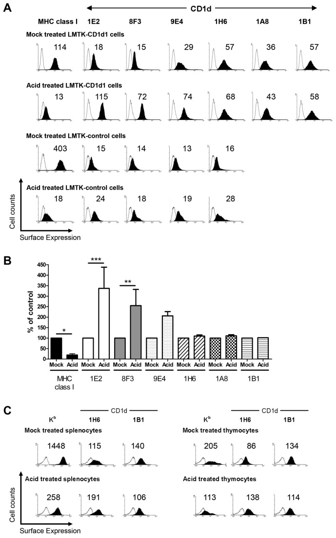
Figure 3. Low pH-mediated disruption of cell surface MHC I molecules enhances cell surface CD1d expression.
(A) Cell surface MHC class I (Kk) and CD1d expression on mock-treated LMTK-CD1d1 (row 1), acid-treated LMTK-CD1d1 (row 2), mock-treated LMTK-control (row 3), and acid-treated LMTK-control (row 4) cells were stained using a pan-murine MHC class I mAb, a panel of mouse CD1d-specific mAbs or their respective isotype control mAb. (B) Changes in MHC class I and CD1d surfaces levels after acid stripping is dependent on the antigen and Ab used. Combined results of multiple experiments with the geometric mean of the mock-treated cells for each mAb normalized to 100; * P < 0.05, ** P < 0.01, *** P < 0.001. (C) Cell surface MHC class I (Kb) and CD1d expression on mock-treated (row 1) and acid-treated (row 2) splenocytes (left) and thymocytes (right) from C57BL/6 mice were measured. The numbers above each histogram indicate the mean fluorescence intensity. Open histogram: isotype control; filled histogram: CD1d- or MHC class I-specific staining.

