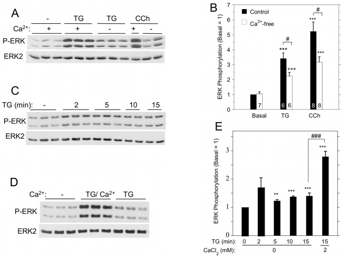
Figure 4. Contribution of extracellular Ca2+ to ERK1/2 activation by TG and carbachol in rat parotid acinar cells.
A. Cells were suspended in the absence or presence (1.8 mM) of Ca2+, and exposed to TG (1 µM) or carbachol (10 µM) for 2 min. B. Quantitative analysis of ERK1/2 phosphorylation in conditions shown in Figure 4A. ***p<0.001 compared to basal, #p<0.05 as indicated. C. Time course of ERK1/2 phosphorylation in cells exposed to TG (1 µM) in the absence of Ca2+. D. Comparison of ERK1/2 phosphorylation in cells in Ca2+-free conditions and exposed to TG (1 µM, 15 min) or exposed to TG (1 µM, 15 min) followed by Ca2+ (1 mM, 2 min) to initiate SOCE. E. Quantitative analysis of ERK1/2 phosphorylation for conditions shown in Figure 4C and 4D. **p<0.01, ***p<0.001 compared to basal; ###p<0.001 as indicated. N = 3–16.

