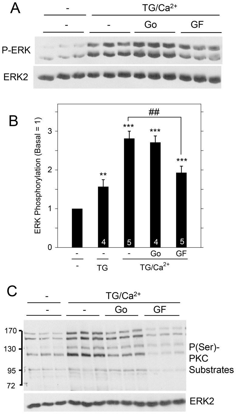
Figure 5. Contribution of PKC proteins to ERK1/2 activation by SOCE in rat parotid acinar cells.
Cells were suspended in the absence of extracellular Ca2+, exposed to TG (1 µM) for 15 min in the absence or presence of GF109203X (10 µM) and Go6976 (1 µM), and then exposed to CaC12 (1 mM, 2 min). A. Effect of PKC inhibitors on ERK1/2 phosphorylation due to Ca2+ entry into cells. B. Quantitative analysis of ERK1/2 phosphorylation for conditions shown in Figure 5A and TG (1 µM, 15 min) alone. **p<0.01, ***p<0.001 compared to basal; ##p<0.01 as indicated. C. Effect of PKC inhibitors in blocking the phosphorylation of PKC substrates in TG-treated cells. Conditions are identical to those shown in Figure 5A. ERK2 was a loading control.

