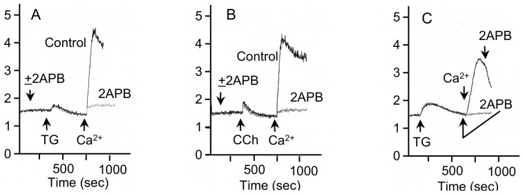
Figure 8. Inhibition of SOCE by 2-APB in rat parotid acinar cells.

A, B. 2-APB (20 µM) blocked the entry of Ca2+ into Fura2-loaded cells in which Ca2+ stores were depleted by the addition of TG (1 µM) (Figure 8A) or carbachol (10 µM) (Figure 8B) to cells in Ca2+-free solution. Vehicle was added to the control cells. C. 2-APB blocks Ca2+ entry after depletion of Ca2+ stores by TG (1 µM) and after the increase in [Ca2+]i produced by addition of 1 mM CaCl2 to store-depleted cells. After depletion of Ca2+ stores and the return of [Ca2+]i to baseline levels, 2-APB (20 µM) was added (gray line) 1 min prior to the addition of 1 mM CaCl2. After the peak increase in Ca2+ produced by addition of 1 mM CaCl2 after store-depletion under control conditions (black line), 2-APB produced an immediate decrease in the level of [Ca2+]i. The ordinate axis is 340/380-nm Fura-2 fluorescence excitation ratio. Shown are individual traces of one experiment, which are representative of at least 3 experiments.
