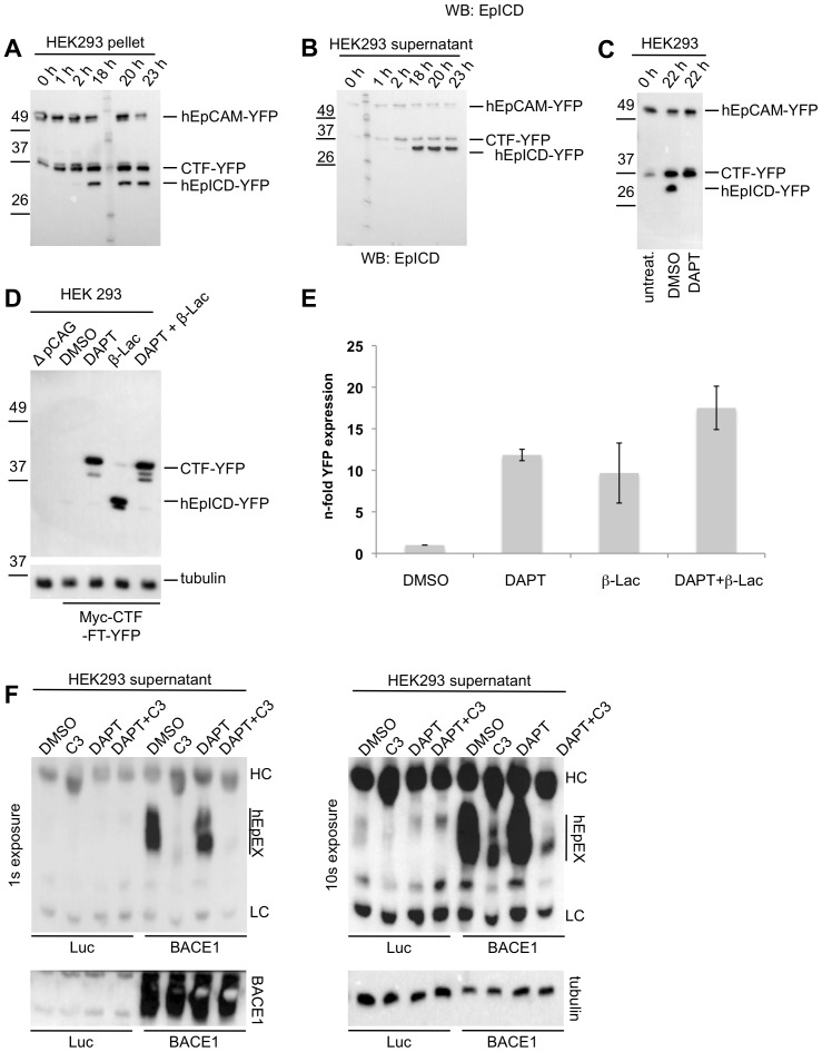
Figure 6. Cleavage and proteasomal degradation of human EpCAM.
HEK293 cells were stably transfected with hEpCAM-YFP and used to determine cleavage products of hEpCAM in membrane assays. Membranes of stable transfectants were isolated and either kept at 0°C (0 h) or incubated at 37°C in reaction buffer for the indicated time points. Thereafter, pellets and supernatant were collected upon differential centrifugation. Pellets (A) and supernatants (B) of membrane assays were separated in a 10% SDS-PAGE and probed with hEpICD- and YFP-specific antibodies. (C) HEK293 hEpCAM-YFP transfectants were treated with DMSO (control) or the γ-secretase inhibitor DAPT before being subjected to a membrane assay. The total fraction of the membrane assay was separated in a 10% SDS-PAGE, and probed with a YFP-specific antibody. Treatment with DAPT resulted in the accumulation of CTF-YFP and in the inhibition of hEpICD-YFP formation. (D) HEK293 human Myc-CTF-TF-YFP transfectants were treated with DMSO (control), the γ-secretase inhibitor DAPT, the proteasome inhibitor β-lacto-lactocystin (β-Lac), or combination of both. Thereafter, whole cell lysates were separated in a 10% SDS-PAGE, and probed with a YFP-specific antibody. Treatment with β-lacto-lactocystin resulted in an accumulation of hEpICD-YFP. Similar loading of protein lysates was visualised upon staining of tubulin on the same blots. Protein bands corresponding to human Myc-CTF-TF-YFP and mEpICD-YFP are indicated in each immunoblot. Shown are the representative results of three independent experiments. (E) YFP fluorescence was analysed in dependency of the treatment of HEK293 human Myc-CTF-TF-YFP transfectants. DMSO treatment served as a reference and values were normalised to one. Shown are the mean values with standard deviations from three independent experiments. (F) HEK293 cells stably expressing hEpCAM-YFP were transiently transfected with expression plasmids for luciferase (Luc) as a control or BACE1 (BACE1). After 24 hours, supernatants were removed and cells treated with the indicated inhibitors of BACE1 (C3), γ-secretase (DAPT) or combinations thereof. After additional 24 hours, supernatants were collected and hEpEX was immunoprecipitated and visualised upon immunoblotting with specific antibodies. Shown are two representative results with exposure times (1 s and 10 s). Over-expression of BACE1 and equal protein loading were verified upon immunobloting (lower left and right panel, respectively).

