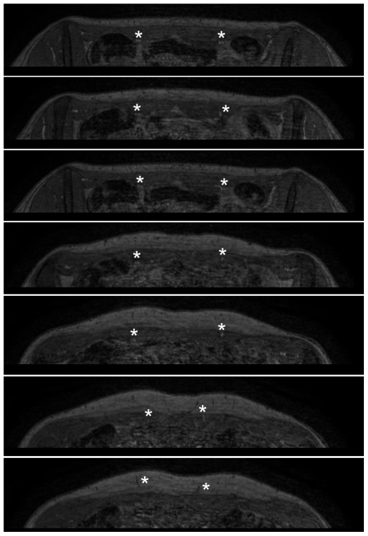
Figure 3. Transverse slices demonstrate the vascular bundle dorsal to the rectus muscle.

Transverse source images of equilibrium-phase dataset in the same patient. Images are from caudal (top panel) to cranial (bottom panel), and clearly demonstrate the vascular bundle dorsal to the rectus muscle shortly after branching off the external iliac artery (asterisks in top three panels). The perforating branches can easily be followed when traversing the rectus muscle to the point where they arise in the subcutaneous fat (asterisks in lower 4 panels).
