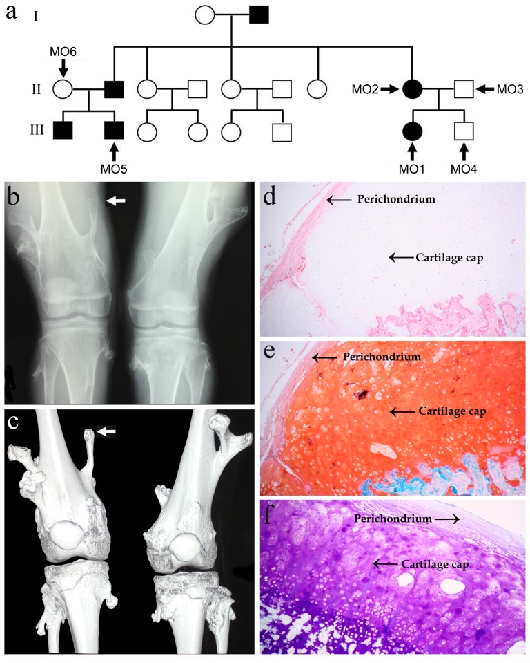
Figure 1. Pedigree structure and characteristic of the MO proband.
(a) Pedigree structure of the MO family; (b,c) computed radiography and 3D reconstruction images of knees of the MO proband. The proband exhibits multiple exostoses, arising from the lateral ends of femurs, tibiae and fibulae. Arrowhead denotes the chondroma used for histochemistry staining; (d–f) low-power micrograph (4×) of the proband’s chondroma sections stained by hematoxylin-eosin (d), Safranin O (e) and Toluidine Blue (f). The cartilage cap of MO is covered by fibrous perichondrium and merges into the underlying spongy bone.

