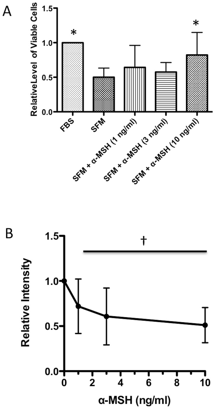
Figure 1. Effects of α-MSH on TUNEL staining of macrophages under serum-free conditions.
The macrophages were incubated for 18 hours under serum-free conditions and treated with α-MSH at 1, 3, and 10 ng/ml. A. The cells were counted by trypan-blue exclusion. The mean relative levels of expected number of viable cells ± SD is presented. * Significance P <0.05 was detected in the cultures treated with 10 ng/ml compared to untreated cells (SFM) based on 8 independent experiments. B. The cells were stained using a TUNEL staining kit, and analyzed by flow cytometry. The mean fluorescent intensity of the TUNEL positive staining was normalized to the mean fluorescent intensity of TUNEL positive staining of the cells cultured under serum free conditions not treated with α-MSH (SFM). † There is a significant (P < 0.05 and Spearman coefficient r = -1) correlation with the decrease in TUNEL staining by increased concentrations of α-MSH treatment. The results are presented as the relative intensities ± SD of 3 independent experiments with the value of 1 for the SFM cells in each experiment.

