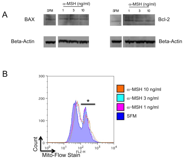
Figure 5. Effects of α-MSH on potential intrinsic mitochondrial pathways of apoptosis.
A. The macrophages were cultured under serum free conditions and were lysed and immunoblotted for BAX and Bcl-2. There was no change in band intensities for BAX and Bcl-2 relative to beta-actin in α-MSH treated and untreated (SFM) cells, nor was there any change in the BAX to Bcl-2 ratio. These results are representative of 3 independent experiments. B. The macrophages were cultured as before, and in the last hour of incubation they were treated with Mito-Flow dye. The cells were assayed by flow cytometry for expression of retained mitochondrial dye in treated and untreated (SFM) macrophages. In all the cultures less than half the cells retained high levels of the dye (*) indicating mitochondrial membrane integrity, with greater than half the cells with low levels of dye indicating loss in mitochondrial membrane integrity. Treating the cells with α-MSH had no effect on this pattern of dye retention. The flow cytometry histogram presented is representative of 3 experiments with the same result of no measurable change seen related to α-MSH treatment. There is no effect on potential intrinsic apoptotic activity by α-MSH.

