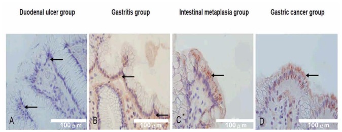
Figure 4. The immunohistochemical stains for integrin α5β1 in the gastric superficial epithelial cells (40X) in the duodenal ulcer group, gastritis group, intestinal metaplasia group, and gastric cancer group, respectively.

The integrin α5β1 was stained on the basolateral membrane of the gastric superficial epithelial cells in the duodenal ulcer group (arrows in A) and gastritis group (arrows in B), but stained on supranuclear or apical surfaces in the intestinal metaplasia group (arrows in C) and gastric cancer group (arrows in D).
