
Figure 2. Inhibition of adhesive strength between IRBC and HDMEC by RGD peptide and anti-β1 mAb.
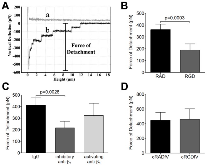
(A) Schematic representation of a typical AFM force curve which depicts a) approach of the IRBC to an endothelial monolayer and b) retraction of the IRBC. The bar indicates the force of detachment that is used as a measure of adhesive strength between IRBC and endothelium. (B) Force measurement on endothelial monolayers pre-incubated with 20 µM RAD or RGD peptide for 30 min at 37°C in 5% CO2 (n = 7). (C) Force measurement on endothelial monolayers pre-incubated with control IgG1, an inhibitory anti-β1 integrin mAb TDM29 or the activating anti-β1 integrin mAb TS2/16 at 10 µg/ml (n = 4). (D) Force measurement on endothelial monolayers pre-incubated with 20 µM cRADfV or cRGDfV peptide (n = 3). For each experiment, 2 IRBC were brought into contact with 3 HDMEC. Contact for 5 min was maintained with a constant force of 150 pN.