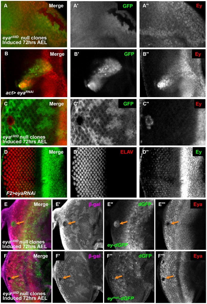
Figure 4. Eya is necessary for ey repression posterior to the morphogenetic furrow.
(A) eyacliIID null clones anterior to the morphogenetic furrow, induced by hs-flp 72 hrs AEL showing GFP and Ey expression (A′) Grayscale image of GFP expression, shown as green in A; loss of GFP expression marks the clones. (A″) Grayscale image of Ey channel alone, red in A. (B) Flipout-Gal4 drives expression of eyaRNAi (VDRC transformant KK108071). Merge of GFP and Ey expression shown. (B′) Grayscale image of GFP expression, shown as green in B; GFP expression marks the clones. (B″) Grayscale image of Ey channel alone, red in B. (C) eyacliIID null clones posterior to the morphogenetic furrow, induced by hs-flp 72 hours after egg lay (AEL) showing GFP and Ey expression (C′) Grayscale image of GFP expression, shown as green in C; loss of GFP expression marks the clones. (C″) Grayscale image of Ey channel alone, red in C. (D) F2-Gal4 drives expression of eyaRNAi (VDRC transformant KK108071). Merge of ELAV and Ey expression shown. (D′) Grayscale image of ELAV expression, shown as green in D, marks differentiating photoreceptors. (D″) Grayscale image of Ey expression, shown as red in D. (E) ey-dGFP (green) expression in eyacliIID null clone posterior to the morphogenetic furrow (indicated by orange arrow) marked by loss of β-Galactosidase expression (magenta) and Eya (red). (E′) Grayscale image of β-Galactosidase expression in E. (E″) Grayscale image of ey-dGFP expression in E as revealed by anti-GFP. (E′″) Grayscale image of Eya expression in E. (F) eymut-dGFP (green) expression in eyacliIID null clone posterior to the morphogenetic furrow (indicated by orange arrow) marked by loss of β-Galactosidase expression (magenta) and Eya (red). (F′) Grayscale image of β-Galactosidase expression in F. (F″) Grayscale image of ey-dGFP expression in F as revealed by anti-GFP. (F′″) Grayscale image of Eya expression in F.

