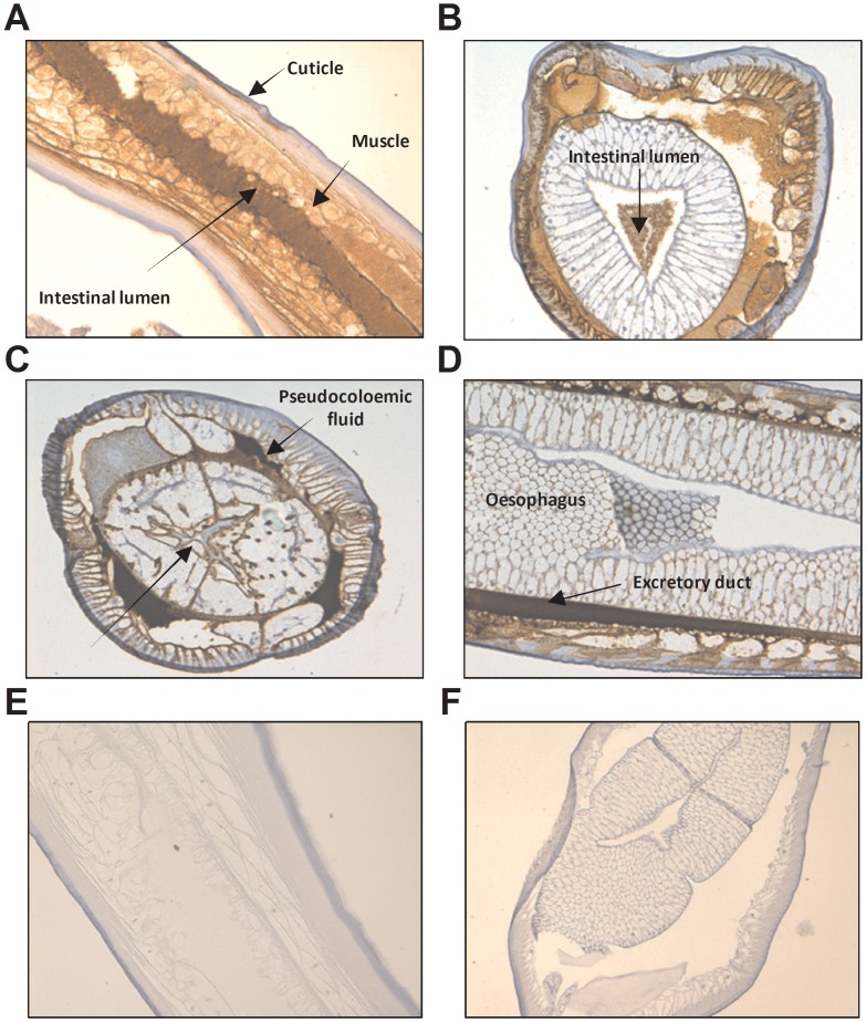
Figure 4. Expression of haemoglobin in Anisakis pegreffii.
A–D) A. pegreffii L3 collected from Thyrsites atun were formalin fixed and stained with 4E8g (Anti-Hb). E–F) A. pegreffii L3 sections were stained with isotype control antibody (mouse IgG1). Detection was carried out using the anti-mouse IgG detection system with DAB. Haemoglobin is stained in brown. Counterstaining was performed with haemotoxylin. All photos taken at 200× magnification.

