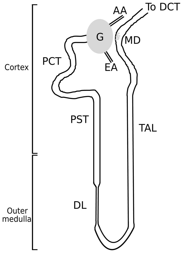
Fig. 1.
A schematic diagram of a short-looped nephron and its renal corpuscle, afferent arteriole (AA), and efferent arteriole (EA). Each nephron consists of a spherical filtering component, the glomerulus (G), and a tubule extending from the renal corpuscle into the proximal convoluted tubule (PCT), proximal straight tubule (PST), and descending limb (DL). Following the loop bend, the thick ascending limb (TAL) rises back into the cortex, then turns into the distal convoluted tubule (DCT). The macula densa (MD) cells at the end of the TAL walls are adjacent to the AA and specialize in sensing the chloride concentration in the downstream fluid. DCT fluid enters the collecting duct system (not shown), where the formation of urine occurs.

