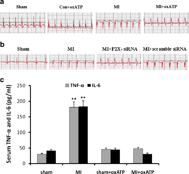
Fig. 1.
Representative traces of ECG in the different groups of rats. Representative traces of ECG were measured 20 days after myocardial ischemia. a The abnormal Q wave appeared obviously in MI rats (n = 8) compared with sham (n = 8) and con+oxATP rats (n = 8). In MI+oxATP group (n = 8), abnormal Q wave induced by MI injury was improved. b After MI rats treated with siRNA P2X7, abnormal Q wave induced by MI injury was improved in comparison with that in MI and MI+scramble siRNA rats. c The serum concentration of TNF-α and IL-6 was measured by ELISA. The concentrations of TNF-α and IL-6 in MI group were higher than those in sham group, con+oxATP group, MI+oxATP group (p < 0.01). No significant difference was found in the concentration of TNF-α or IL-6 among sham group, con+oxATP group, and MI+oxATP group (p > 0.05). The n value is 8 in all groups. Results are mean ± SE. **p < 0.05 vs sham group, con+oxATP group, and MI+oxATP group

