
Fig. 2.
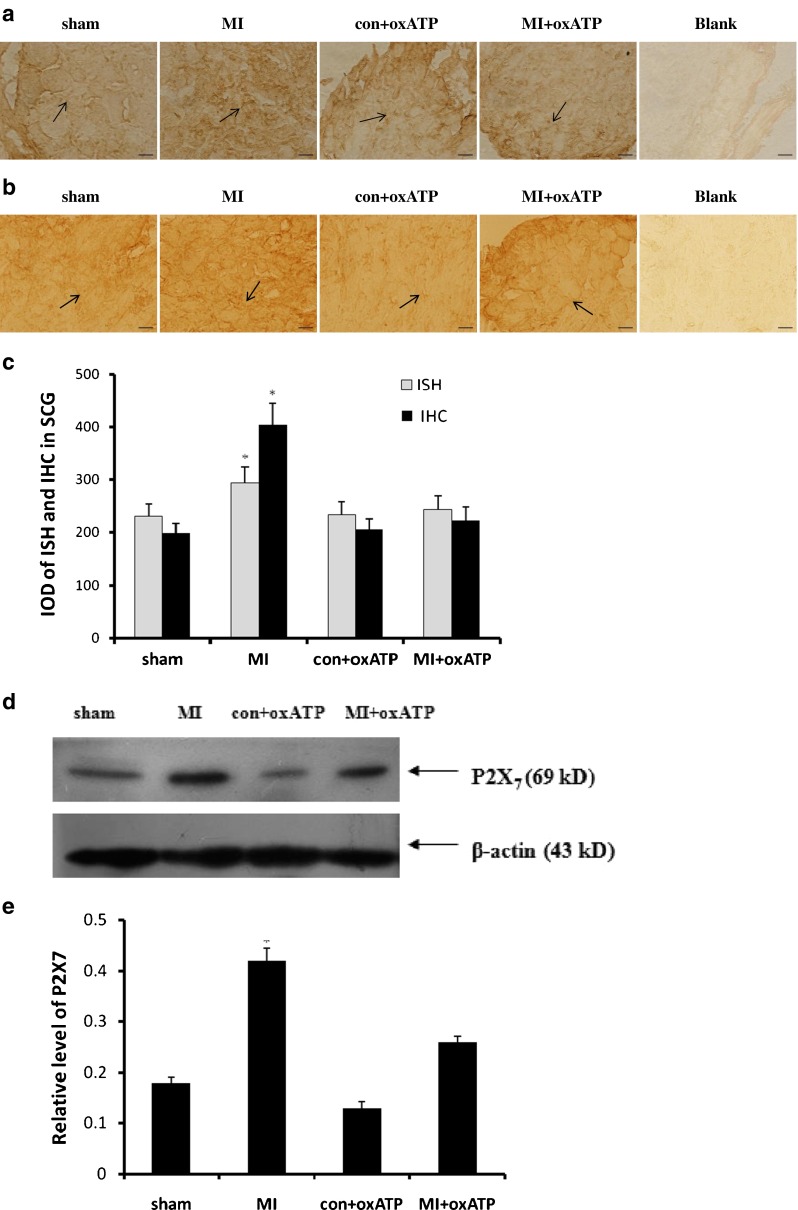
Effects of oxATP on the increased P2X7 expression induced by ischemic injury. The expressions of P2X7 mRNA and protein were measured by in situ hybridization (a), immunohistochemistry (b), and Western blotting (d). The bar graphs (c, e) showed the statistical results for expression of P2X7 mRNA or protein. The relative expression in Western blotting was expressed by the IOD ratio of P2X7 protein to β-actin. The results showed that the expression levels of P2X7 mRNA or protein in MI group (n = 8) were significantly higher than those in sham group (n = 8), con+oxATP group (n = 8), and MI+oxATP group (n = 8; p < 0.05). No difference was found among sham group, con+oxATP group, and MI+oxATP group (p > 0.05). Arrows indicate the immunostaining neurons. Scale bars, 20 μm. Results are mean ± SE. *p < 0.05 vs sham group, con+oxATP group, and MI+oxATP group