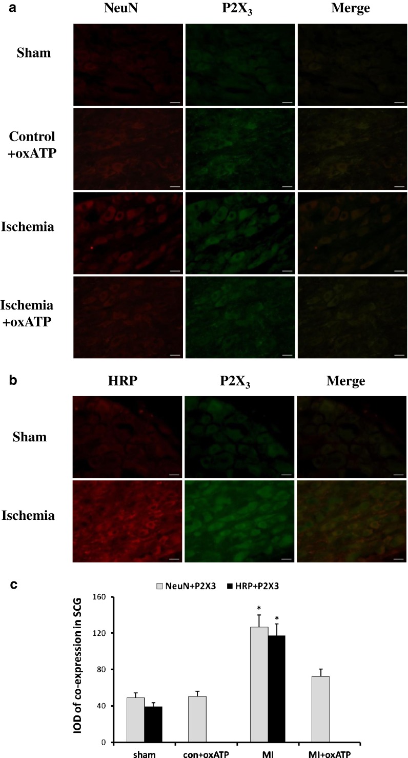
Fig. 6.
The double-label immunofluorescence of P2X3 and NeuN or HRP in SCG. a NeuN is the marker of neurons. The image shows that P2X3 receptor was mainly expressed in SCG neurons. Red signal represents P2X3 staining with TRITC, and green signal indicates NeuN staining with FITC. Merge represents P2X3 and NeuN double staining image. Scale bar, 20 μm. b There was double immunofluorescence of P2X3 and HRP from the cardiac afferent endings to SCG, as tested by the retrograde neuronal labeling. Red signal represents P2X3 staining with TRITC, and green signal indicates HRP staining with FITC. Merge represents P2X3 and HRP double staining image. Scale bar, 20 μm. c The bar graphs showed the statistical results for co-expression of P2X3 and NeuN or HRP in SCG (n = 6 in all groups)

