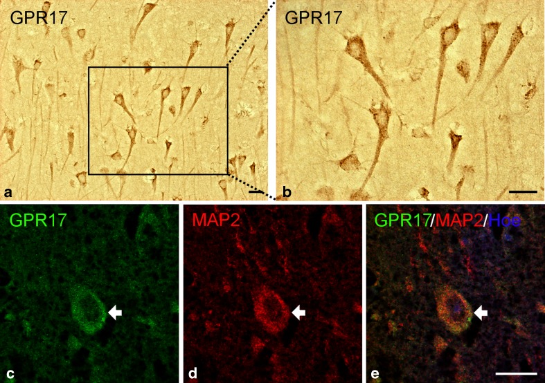
Fig. 5.
GPR17 expression in neurons after TBI. Immunohistochemical analysis of tissues collected from human autoptic TBI samples revealed expression of GPR17 in cells resembling pyramidal neurons (a) and higher magnification detail (b). Immunofluorescence staining confirmed the neuronal nature of these cells: green fluorescence for GPR17 (c); red fluorescence for the neuronal marker MAP2 (d); merge of the two images showing coexpression of GPR17 and MAP2 in the same cell (e). Nuclei are labeled with Hoechst 33258 dye (Hoe, blue fluorescence). Scale bars = 20 μm (a), 50 μm (b), and 20 μm (c–e)

