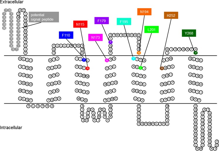
Fig. 1.
Topology model of the rat adenine receptor. Mutated amino acid residues presumed to be involved in ligand binding and the potential N-terminal signal peptide are shown as hexagons using the following color code: gray, potential signal peptide; blue, Phe1103.24; red, Asn1153.29; magenta, Asn1734.60; purple, Phe17945.39 (Phe179 is located in ECL2 which also includes the highly conserved residue Cys19045.50); orange, Asn1945.40; cyan, Phe1955.41; green, Leu2015.47; brown, His2526.54; and dark green, Tyr2687.32

