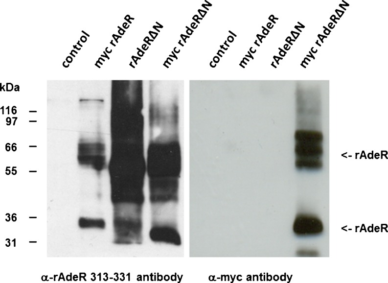
Fig. 9.
Western blot experiments were performed with membranes of Sf9 insect cells (30 μg of protein, control cells) and membranes containing myc-rAdeR, rAdeRΔN, or myc-rAdeRΔN, respectively (50 μg of protein each). α-myc antibody recognizes the myc epitope; α-rAdeR-313-331 antibody recognizes the rAdeR peptide (amino acid residues 323–331). The protein bands were detected at 35 kDa. Additional rAdeR-specific bands were detected between about 60 and 70 kDa possibly representing receptor dimers

