
FIGURE 4.
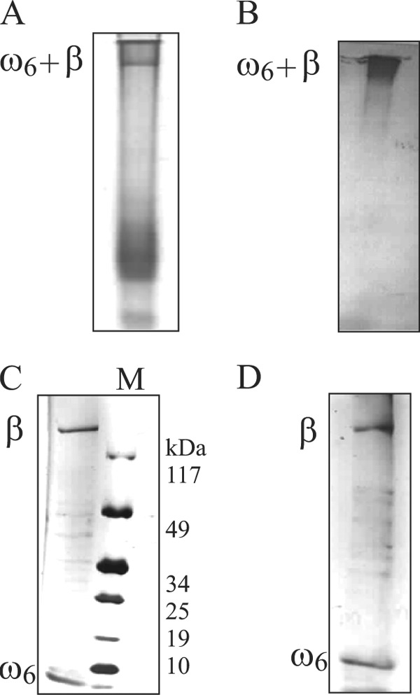
ω6-β′ association studies. Shown are 8–20% gradient SDS-PAGE of the ω6-β′ (A) and the position of ω6-β′ complex in A inferred by immunoblotting with anti-ω antibody (B). The gel slice bearing ω6-β′ complex in A was excised, and its contents were electrophoresed on an 8–20% gradient SDS-polyacrylamide gel (C), and the constituents of the preformed ω6-β′ complex were identified by immunoblotting with anti-ω and anti-β′ antibodies (D). M denotes the molecular weight marker.