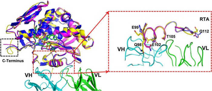
FIGURE 3.

Superposition of RTA in the free form (PDB code 2AAI, yellow), in the complex with 6C2 (PDB code 4KUC, magenta) and in the complex with cyclic G (9-DA) GA2′-OMe (PDB code 3HIO, blue). Cyclic G (9-DA) GA2′-OMe (which contains the tetranucleotide sequence of the GAGA sarcin-ricin loop but with the ricin-susceptible adenosine replaced with 4′-deaza-1′-aza-2′-deoxy-1′-(9-methylene)-immucillin-A (9-DA), a transition state mimic) serves to mimic the substrate and is shown as sticks (green). 6C2 is colored the same as described in the legend to Fig. 1A. The overall structures are almost identical. Changing of the residues on the epitope is highlighted by the red dashed lines.
