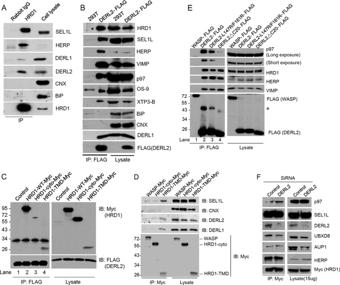
FIGURE 3.
DERL2 associates with HRD1-SEL1L complex requiring TMD of HRD1. A, 293T cells were lysed in HKM buffer with 1% deoxy-bigChap and protease inhibitors and immunoprecipitated (IP) with HRD1 antibodies. The precipitates were separated in SDS-PAGE and immunoblotted (IB) with the indicated antibodies. B is as in A but with 293T cells stably expressing DERL2-FLAG and immunoprecipitated with FLAG-M2 beads. Immunoblotting was carried out with the indicated antibodies. C, Myc-tagged HRD1 expression plasmids were transfected to 293T cells stably expressing DERL2-FLAG. The cells were lysed in HKM buffer with protease inhibitors and 1% digitonin and immunoprecipitated by FLAG-M2 beads. Precipitates were separated on SDS-PAGE and immunoblotted with FLAG or Myc antibodies. D, Myc-tagged HRD1 or WASP plasmids were transfected to 293T cells. The cells were lysed in HKM buffer with 1% Triton X-100 and protease inhibitors, immunoprecipitated by Myc antibodies, and immunoblotted with Myc, SEL1L, calnexin (CNX), DERL1, and DERL2 antibodies. E, 293T cells were transfected with various FLAG-tagged DERL2 plasmids as indicated. Cells were lysed, and immunoprecipitation was carried out with FLAG-M2 beads. Precipitates were separated on SDS-PAGE and immunoblotted with the indicated antibodies. The asterisk (*) marks the dimeric DERL2. F, 293T cells stably expressing Myc-HRD1 proteins were depleted of DERL2 by siRNA oligonucleotides. The cells were lysed and immunoprecipitated using Myc antibodies in HKM buffer containing 1% deoxy-bigChap. The immunoprecipitate and cell lysates were separated on SDS-PAGE and immunoblotted with the indicated antibodies.

