FIGURE 5.
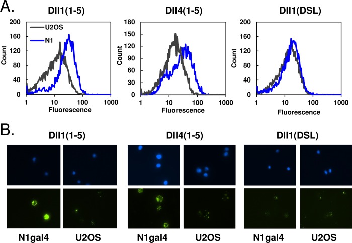
Ligand binding to Notch cells. A, detection of purified ligands binding to Notch-bearing cells. Biotinylated ligands were coupled to yellow fluorescent avidin beads and incubated with Notch1-gal4 U2OS or U2OS control cells. When analyzed via flow cytometry, the N1gal4 cells incubated with Dll1(1–5) and Dll4(1–5) showed a shift in fluorescence relative to U2OS control cells, which was not observed with the shorter Dll1(DSL) ligand. B, visualization of ligand-cell binding with fluorescence microscopy. Top and bottom rows show DAPI and FITC staining, respectively. Beads coated with Dll1(1–5) or Dll4(1–5) both bound preferentially to Notch-bearing cells over control cells, whereas Dll1(DSL)-coated beads did not.

