
FIGURE 1.
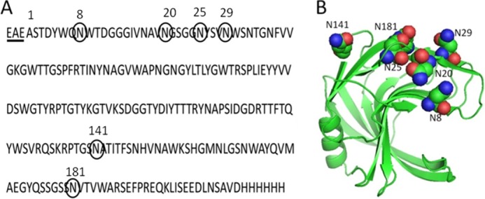
Amino acid sequence (A) and ribbon representation (B) of the crystal structure of xylanase A from B. subtilis showing the potential N-glycosylation sites. The sequence was modified to contain a C-terminal peptide with a polyhistidine. The underlined residues (EAE) are derived from the α-factor signal peptide. The numbers of amino acids correspond to the native sequence that does not include residues EAE.