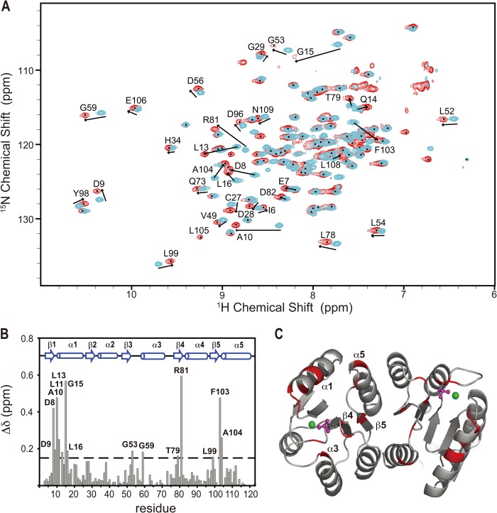
FIGURE 3.
The induced chemical shift perturbation of 2H,15N,13C-labeled PmrAN with the presence of PmrD. A, a section of overlaid 1H,15N TROSY-HSQC spectra of free PmrAN (cyan) binding to 2-fold molar ratio of unlabeled PmrD (red). The residues showing significant chemical shift perturbations are labeled with a one-letter code for amino acids with the residue number. B, graph representing the weighted average of the chemical shift differences between the free and PmrD-bound PmrAN. The secondary structural elements of PmrAN are illustrated on top. C, ribbon structure of PmrAN with the residues showing weighted chemical shift perturbations > 0.135 ppm are in red.

