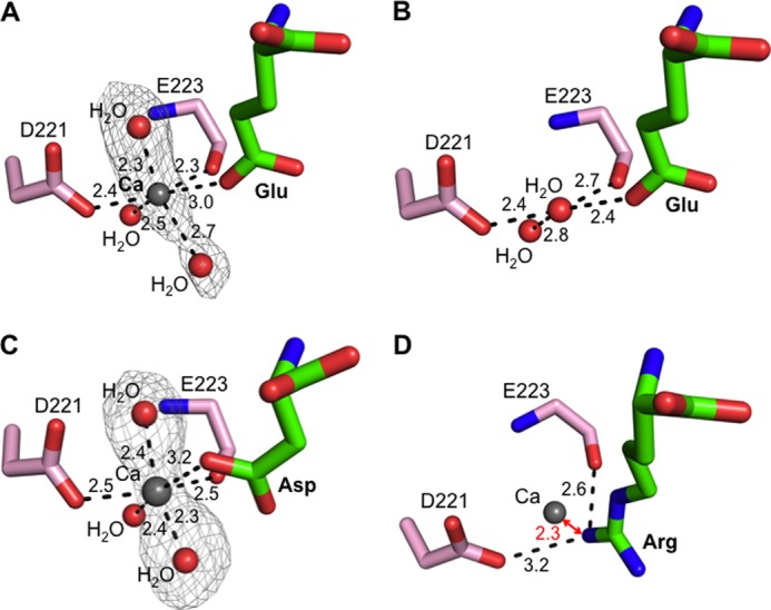
FIGURE 7.

Calcium-modulated substrate specificity of APA. A, observed calcium-binding site in the S1 pocket of glutamate-bound APA. The electron density map of calcium and two additional calcium-coordinating water molecules corresponds to the Fo − Fc omit map (contoured at 3.5σ) that was calculated with a water molecule occupying the calcium-binding site and in the absence of the two additional water molecules. B, in the absence of calcium, a water molecule occupies the calcium-binding site in the S1 pocket of glutamate-bound APA. C, observed calcium-binding site in the S1 pocket of aspartate-bound APA. The electron density map was calculated in the same way as described for A, except that it was contoured at 3.0σ. D, modeled calcium-binding site in the S1 pocket of arginine-bound APA. In the presence of calcium, arginine was not observed upon soaking into APA crystals. Instead, a structural model was constructed in which calcium replaced the water molecule occupying the calcium-binding site as in Fig. 5B.
