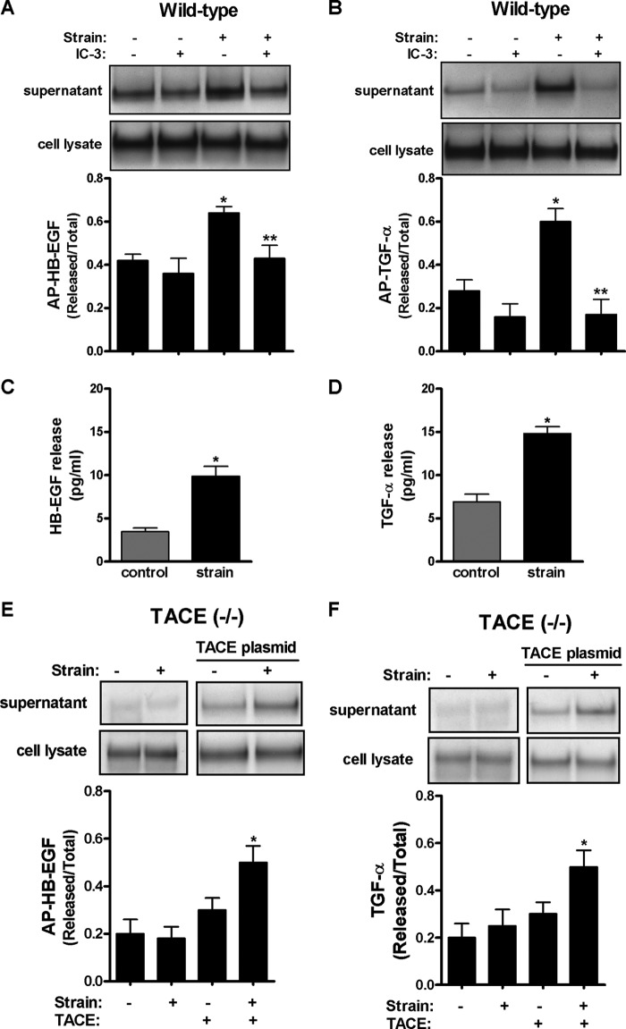
FIGURE 2.
Strain-induced release of HB-EGF and TGF-α is mediated via TACE. A and B, E17 type II cells were transfected by electroporation with plasmids encoding alkaline phosphatase (AP)-tagged HB-EGF (A) or TGF-α (B) ligands in the presence or absence of IC-3 (10 μm), a TACE inhibitor, and then exposed to 5% cyclic strain for 30 min. Samples were processed to assess HB-EGF or TGF-α shedding as described in “Experimental Procedures.” n = 4, *, p < 0.05 versus control; **, p < 0.01 versus strain without inhibitor. C and D, E17 type II cells were exposed to 5% cyclic strain for 30 min. Supernatants were collected, concentrated and processed to detect shedding of mature HB-EGF (R & D, cat. DY259) and TGF-α (R & D, cat. DY239) by ELISA following manufacturer's recommendations. Samples were normalized to the concentration of proteins in the cell lysate. p < 0.05 versus control. Results are from three independent experiments. E and F, E17 type II cells isolated from TACE knock-out mice and transfected or not with a plasmid encoding the full-length of TACE were exposed to similar experimental conditions as described above to investigate shedding of mature HB-EGF (E) or TGF- α (F). n = 4, *, p < 0.05 versus control plus TACE transfection. Upper panels show representative blots.

