
FIGURE 3.
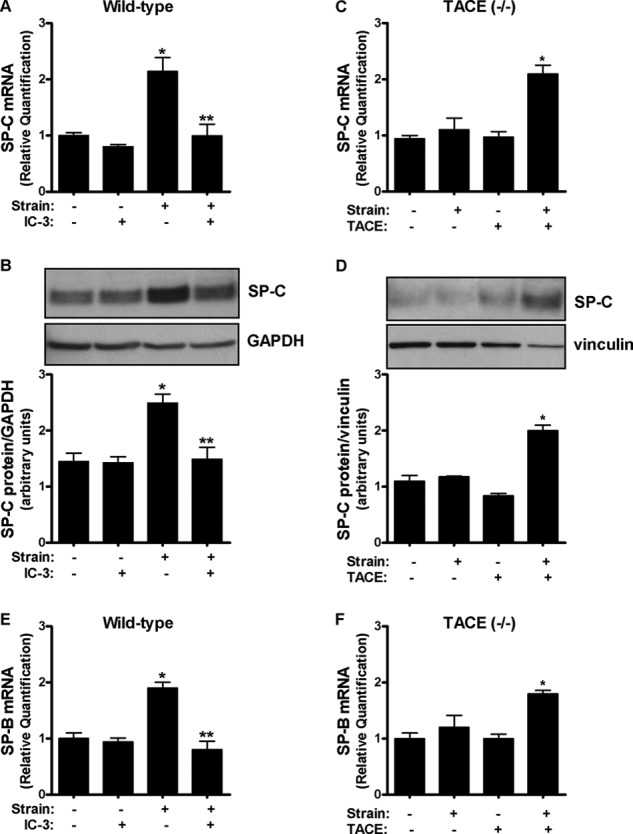
Strain-induced fetal type II cell differentiation is mediated via TACE. A and B, E17 type II cells were exposed to 5% cyclic strain for 24 h in the presence or not of IC-3 (10 μm), a TACE inhibitor. Samples were analyzed by qRT-PCR (A) or Western blot (B) to detect SP-C abundance. n = 4, *, p < 0.05 versus control; **, p < 0.01 versus strain without IC-3. C and D, type II cells isolated from TACE knock-out mice were transfected or not with a plasmid encoding the full-length of TACE and then exposed to similar experimental conditions as described in A and B. n = 4, *, p < 0.05 versus control plus TACE transfection. Upper panels in B and D show representative blots. E and F, E17 cells were exposed to similar experimental conditions described in A and C but SP-B, instead of SP-C, was used as a marker of differentiation.