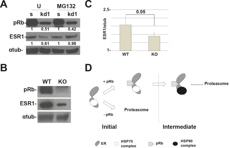
Figure 5. RB1 kd primary human mammary cells and Rb1 KO mice show a reduction in ESR1 protein levels.
(A) HMEpC RB1 kd1 cells express 50% less of the ESR1 protein compared to scrambled cells. Treatment with MG132 for 4 hours rescued the expression of the ESR1 protein. The quantification represents the ratio between the ESR1 and alpha-tubulin proteins and normalized to scrambled cells. (B) Rb1 conditional KO mice in mammary cells have a reduced expression of ESR1. (C) Quantification of 6 mice as in (B). (D) Working model of pRb and the chaperone complex that regulate ESR1 turnover. After the assembly of the initiation complex, pRb is necessary for the recruitment of the HSP90 complex that forms the intermediate complex. Without pRb, the ESR1 is more ubiquitinated and degraded by proteasome pathway.

