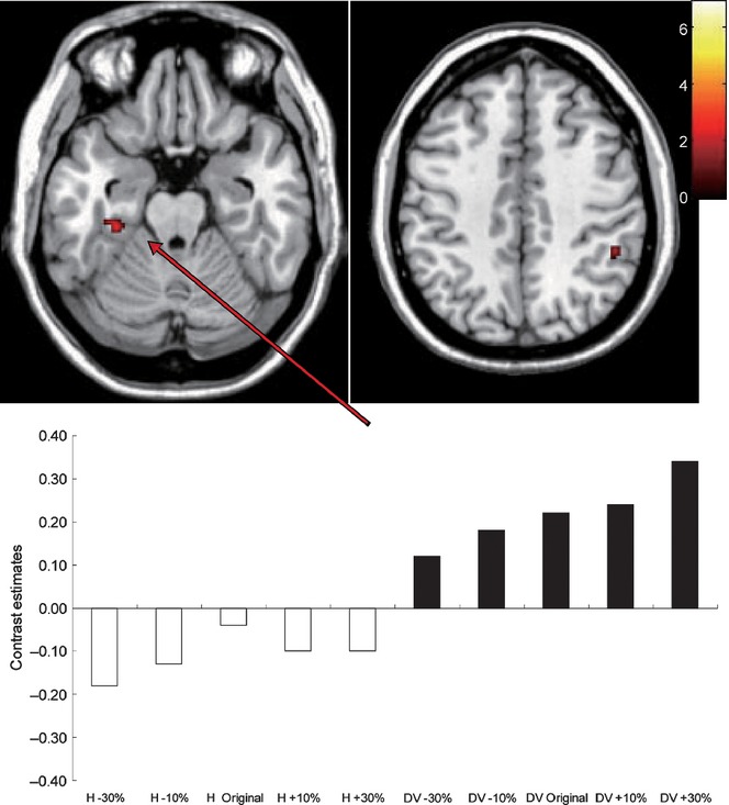
FIG. 3.

The left fusiform body area (FBA, left) and the right inferior parietal lobule (IPL; right) exhibited greater responses to body images of dorsoventral thickness than those of horizontal width. In the bottom panel, the contrast estimates for the FBA (MNI coordinates of x, y, z: −32, −28, −22; or a nearest peak) corresponding to each body image are shown. The color bar shows the t-value.
