Fig. 3.
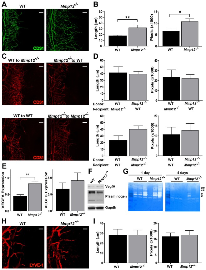
MMP12 alters corneal angiogenesis following injury. (A) Micrographs of whole-mount corneal preparations stained for angiogenic endothelium (CD31) 6 days after injury. (B) Quantitative analysis of angiogenesis, showing lengths of individual vessels in injured Mmp12−/− corneas were 1.7 times longer compared with those in WT corneas (32±4.9 µm versus 18±1.2 µm; n = 11, P<0.05). Additional morphometric comparison of the area covered by CD31+ vessels in WT and Mmp12−/− corneas confirmed that the area was larger in the Mmp12−/− corneas as compared with that in WT control mice (10738±1245 pixels versus 6513±1017 pixels; n = 11, P<0.05). (C) Micrographs demonstrating corneal angiogenesis 6 days after injury in mice that underwent bone marrow transplantation. (D) Quantitative analysis of angiogenesis following bone marrow transplantation. After transplantation of WT and Mmp12−/− bone marrow cells into Mmp12−/− and WT recipient mice, respectively, neovascular lengths and areas were similar (41±8.9 µm versus 39±4.0 µm and 23273±7415 pixels versus 21850±4104 pixels, n = 7). Transplantation of Mmp12−/− bone marrow cells into Mmp12−/− recipient mice resulted in increased neovascular lengths compared with transplantation of WT bone marrow cells into WT donor mice (40±6.8 µm versus 23±6.8 µm and 12759±3455 pixels versus 10663±3698 pixels; n = 3, P<0.05). (E) Quantification of VEGFA and VEGFB mRNA expression levels in WT and Mmp12−/− mouse corneas 6 days after corneal injury. Results are means ± s.e.m., n = 8, **P<0.005. (F) Levels of VEGFA and plasminogen cleavage products in corneal lysates at 6 days post-injury. Western blots were probed with GAPDH as a loading control. (G) Corneal tissues at 1 and 4 days post-injury were homogenized and soluble lysates (10 µg per well) were subjected to gelatin zymography. Protein standards were used to identify inactive MMP on zymograms and are indicated with asterisks (**MMP9 and *MMP2). (H) Representative whole-mount micrographs stained for lymphangiogenic (LYVE-1) endothelium 6 days after injury. (I) Quantitative analysis of lymphangiogenesis, measured as vessel lengths pixel density. Values are means ± s.e.m. (n = 11). Scale bars: 10 µm.

