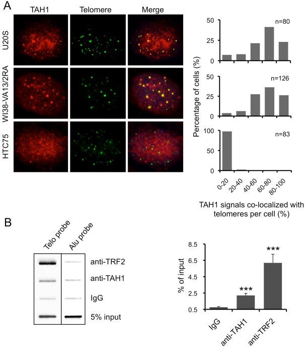
Fig. 1.
TAH1 localizes to telomeres more frequently in ALT cells than in telomerase-positive cells. (A) IF-FISH was carried out for endogenous TAH1 using the telomere PNA-TelC-FITC probe in U2OS, WI38-VA13/2RA and HTC75 cells. Quantifications of the percentage of cells containing varying degrees of TAH1–telomere co-localization are shown on the right. (B) Telomere ChIP analysis with the biotinylated telomere probe (TTAGGG)3 was performed using U2OS cells and anti-TAH1 antibodies. IgG served as IP control and an Alu probe was used for a loading control. A quantification of the data is shown on the right. For TRF2, n = 3. For TAH1 and IgG, n = 6. Error bars indicate standard error. ***P<0.001 compared with control.

